
Research
Projects
Medical and Pharmaceutical Applications of Secondary Ion Mass Spectrometry
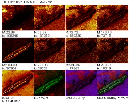
Identification and localization of biomolecules in cells and tissue samples are important for understanding of subcellular structures. Isolated cardiac cells and tissue of rats are studied by using secondary ion mass spectrometry, providing chemical composition of cardiac cell membrane and tissue surface in native form. The result is a spatially resolved chemical imaging as a lateral distribution of biologically relevant molecules—phospholipids, along with fatty acids, and cholesterol. Phospholipids are represented by phosphatidylcholine and cardiolipin molecules and their fragments. Phosphatidylcholine polar head group at mass of 184.1 u has an origin in the cell membrane, and a two-dimensional distribution of this fragment provides clear chemical contours of the cell. The high-resolution contrast of the cell is observed within its environment represented with Na+ ions. Images of PO4H- fragment and fatty acids with 16 or 18 C atoms are determined in cardiac tissue. Distributions of these 16 and 18 C fatty acids are the same within their groups, and interestingly, these two distribution groups are spatially complementary. Contours of phosphatidylcholine and cardiolipin fragments are also complementary, the distributions of 16 C fatty acids and phosphatidylcholine are identical, and the distributions of 18 C fatty acids and cardiolipin are also the same. The secondary ion mass spectrometry is the excellent tool also for idetification of trace elements, like Si and Al, or Sr as part of the supplement, being helpful in medical and pharmaceutical research.
- Jerigova, Monika - Chorvatova, Alzbeta - Chorvat, Dusan - Biro, Csaba - Velic, Dusan: Analysis of cardiac tissue by gold cluster ion bombardment, Applied Surface Science. - Vol. 252, No. 19, Sp. Iss. (2006), s. 6782-6785
- Jerigova, Monika - Michalka, Miroslav - Kopani, Martin - Rychly, Boris - Jakubovsky, Jan - Velic, Dusan: Microscopy and chemical imaging of Behcet brain tissue, Applied Surface Science. - Vol. 255, No. 4 (2008), s.1584-1587
- Jerigova, Monika - Biro, Csaba - Kirchnerova, Jana - Chorvatova, Alzbeta - Chorvat, Dusan - Lorenc, Dusan - Velic, Dusan: Chemical Imaging of Cardiac Cell and Tissue by Using Secondary Ion Mass Spectrometry, Molecular Imaging and Biology. - Vol. 13, No. 6 (2011), s. 1067-1076
- Jerigova, Monika - Stancikova, Maria - Rovensky, Jozef - Velic, Dusan: Strontium distribution in bones and tissues of strontium ranelate-administrated rats, Surface and Interface Analysis. - Vol. 43, No. 1-2 (2011), s. 306-309
- Stupavska, Monika - Jerigova, Monika - Velic, Dusan: Matrix and primary ion-related aspects of tryptophan SIMS analysis, Surface and Interface Analysis. - Vol. 45, No. 1 Sp. Iss. (2013), s. 68-71
- Stupavska, Monika - Jerigova, Monika - Velic, Dusan: Alkaline earth metal salts of CaCO3, BaCO3, and SrCO3 as matrix for tryptophan SIMS analysis, Surface and Interface Analysis. - Vol. 43, No. 1-2 (2011), s. 462-466